
How Do Peptides Work? 7 Essential Mechanisms in 2026
Peptides work through receptor binding and signal transduction cascades. Explore 7 essential mechanisms, synthesis, delivery challenges, and research storage in 2026.
📋 Table of Contents
Peptides work by binding to specific receptors on cell surfaces, triggering intracellular signal transduction cascades that alter gene expression, enzyme activity, or ion flow. In laboratory research, understanding how do peptides work requires examining amino acid sequence, receptor affinity, and downstream second messengers such as cAMP, Ca2+, and IP3. This research-use-only overview explains peptide mechanisms, delivery challenges, and practical considerations for investigators sourcing research-grade peptides.
⚠️ Research Use Only Notice: The products discussed on this site are sold strictly for in vitro laboratory research purposes only. They are not for human use, not for veterinary use, and not intended to diagnose, treat, cure, or prevent any disease. All information presented is for educational purposes in a research context. Must be 21+ to purchase.
Key takeaways
- Peptides are short chains of amino acids, typically 2–50 residues, that act as ligands for receptors such as GPCRs.
- How do peptides work depends on receptor binding affinity, conformational change, and intracellular signal transduction.
- Second messengers (cAMP, Ca2+, IP3) amplify peptide signals inside cells.
- Peptide stability and bioavailability remain central delivery challenges; PEGylation and cyclization are common research modifications.
- Research-grade peptides require verified purity, sequence confirmation, and proper storage.
What Are Peptides? Amino Acid Chains vs. Proteins
Peptides are linear or cyclic chains of amino acids linked by peptide bonds. By convention, chains shorter than roughly 50 amino acids are called peptides; longer chains are proteins. This size boundary is not absolute, but it helps researchers classify molecules by folding complexity, stability, and receptor interactions. Common peptide hormones such as insulin (51 amino acids) and oxytocin (9 amino acids) illustrate how sequence length directly influences receptor binding and biological activity.
In laboratory settings, researchers synthesize peptides with defined sequences to probe receptor pharmacology. The amino acid sequence determines secondary structure, charge distribution, and hydrogen-bonding potential. Because peptides are smaller than proteins, they often lack stable tertiary folds, which affects how do peptides work in solution and at membrane interfaces. This structural flexibility can be an advantage for receptor docking but a disadvantage for proteolytic stability.
How Do Peptides Work as Signaling Molecules? Lock-and-Key Receptor Binding
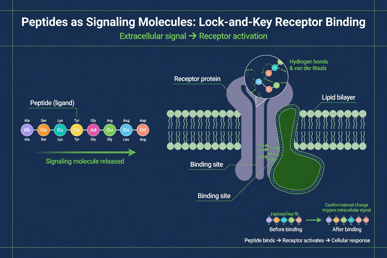
At the molecular level, how do peptides work through a lock-and-key interaction with cell-surface or intracellular receptors. A peptide ligand binds to a complementary binding pocket on its receptor, inducing a conformational change. This change activates the receptor’s intracellular domain, initiating downstream signaling. The specificity of this binding depends on hydrogen bonds, hydrophobic contacts, and electrostatic interactions between peptide side chains and receptor residues.
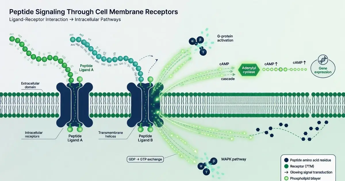
Most peptide receptors belong to the G protein-coupled receptor (GPCR) superfamily. GPCRs span the membrane seven times and couple to heterotrimeric G proteins. When a peptide ligand binds, the receptor exchanges GDP for GTP on the Gα subunit, causing dissociation of Gα and Gβγ. These subunits then regulate effector enzymes such as adenylyl cyclase and phospholipase C. This is the core answer to how do peptides work in many endocrine and neuroendocrine systems.
Why Does Receptor Subtype Determine How Peptides Work?
Peptide receptors include GPCRs, receptor tyrosine kinases, and guanylyl cyclase-coupled receptors. Each receptor class uses distinct intracellular domains to translate extracellular binding into cellular responses. For GPCRs, ligand specificity arises from the shape and charge of the orthosteric binding pocket. For example, oxytocin and vasopressin receptors share high sequence homology but discriminate between their peptide ligands through subtle differences in transmembrane residues. This discrimination is a key variable when researchers ask how do peptides work across related receptor subtypes.
💡 Key Takeaway: How Do Peptides Work as Signaling Molecules? Lock-and-Key Receptor Binding
How Do Signal Transduction Cascades Amplify Peptide Effects?
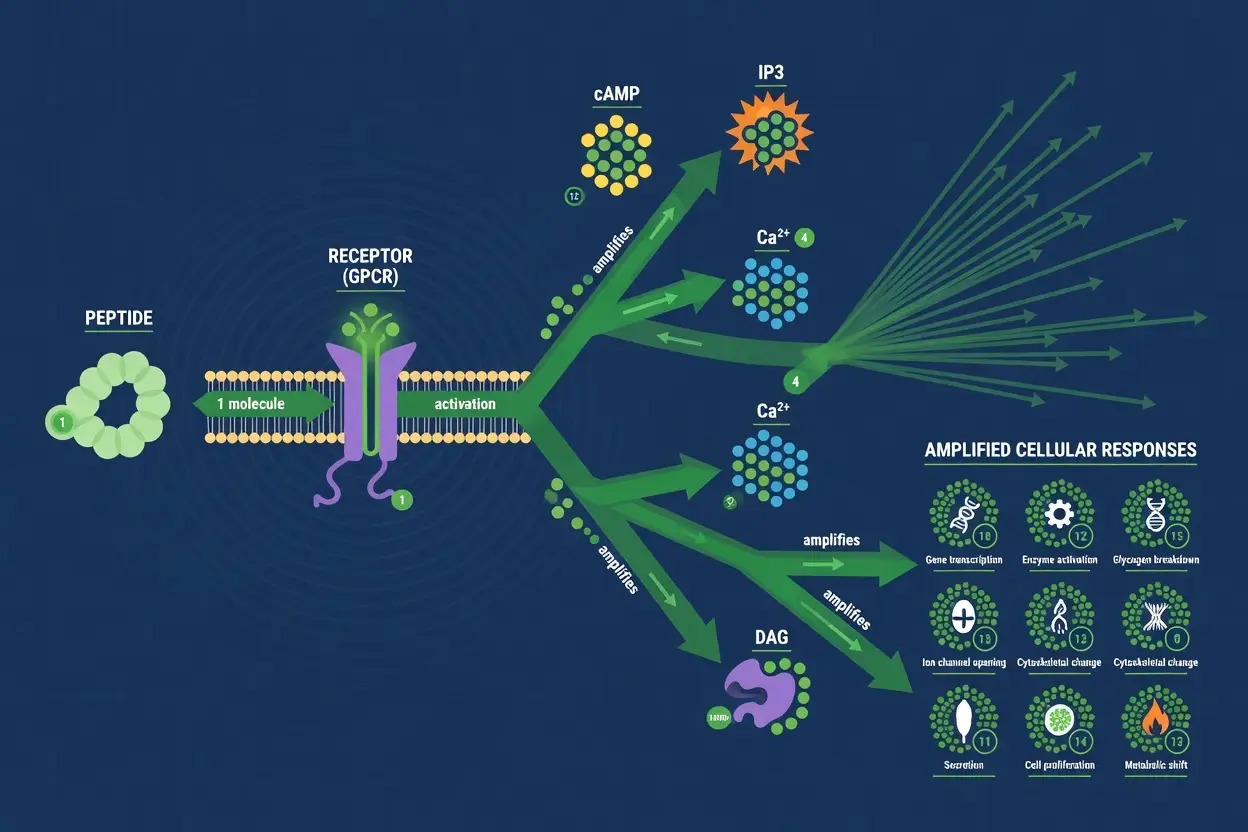
Once a peptide receptor is activated, signal transduction cascades amplify the extracellular signal. For GPCRs coupled to Gαs, adenylyl cyclase converts ATP to cyclic AMP (cAMP). cAMP activates protein kinase A (PKA), which phosphorylates target proteins including transcription factors. For Gαq-coupled receptors, phospholipase C cleaves PIP2 into IP3 and diacylglycerol (DAG). IP3 releases Ca2+ from the endoplasmic reticulum, while DAG activates protein kinase C (PKC). These second messengers—cAMP, Ca2+, and IP3—are central to understanding how do peptides work at the intracellular level.
Peptide signaling can also proceed through receptor tyrosine kinases (RTKs), although fewer peptides use this route. Insulin, for example, binds the insulin receptor, a dimeric RTK, leading to autophosphorylation and recruitment of IRS adaptor proteins. This activates PI3K/Akt and MAPK pathways. Oxytocin, by contrast, acts through a GPCR coupled to Gαq, raising intracellular Ca2+ in smooth muscle and neuronal cells. Comparing these two peptide hormones shows that how do peptides work is not a single mechanism but a family of receptor-specific pathways.
Signal termination is equally important. Phosphodiesterases degrade cAMP, inositol phosphatases dephosphorylate IP3, and Ca2+ ATPases restore cytosolic Ca2+ levels. Receptor desensitization occurs via GPCR kinases and β-arrestin recruitment. These negative feedback loops ensure that peptide signals are transient and tightly controlled. In experimental systems, blocking these termination steps can reveal how do peptides work under sustained versus pulsatile stimulation. For a detailed review of GPCR signaling, see PubMed: Peptide receptor signaling and NIH Bookshelf: Signal Transduction.
What Are Real-World Peptide Functions? Hormones, Repair, and Immunity
Endogenous peptides regulate diverse physiological processes. Insulin controls glucose uptake; oxytocin modulates social bonding and uterine contraction; glucagon-like peptide-1 (GLP-1) regulates insulin secretion; antimicrobial peptides disrupt microbial membranes. In research, these peptides serve as reference standards for receptor binding assays and as templates for synthetic analogs. The question how do peptides work in these contexts often reduces to receptor subtype expression and downstream effector coupling.
Peptides involved in tissue repair include thymosin beta-4, BPC-157, and GHK-Cu, which are studied in cell culture and animal models for their effects on migration, proliferation, and extracellular matrix deposition. However, no therapeutic claims are made here. Research-use-only peptides are supplied for in vitro assays, receptor binding studies, and preclinical investigations. Investigators must verify purity, sequence, and biological activity before use. For a broader overview of research considerations, see peptides benefits and risks — 7 Proven Facts for 2026.
Immunomodulatory peptides such as thymosin alpha-1 and LL-37 are studied for receptor-mediated effects on immune cells. These peptides bind receptors on macrophages, dendritic cells, and lymphocytes, modulating cytokine release and phagocytosis. Understanding how do peptides work in immune cell assays requires careful control of endotoxin levels, because even trace lipopolysaccharide can confound results.
How Are Research Peptides Synthesized and Verified?
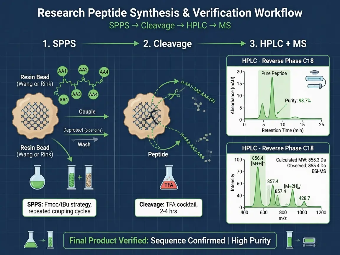
Solid-phase peptide synthesis (SPPS) is the standard method for producing research peptides. The process builds the peptide chain from the C-terminus to the N-terminus on a resin support, using Fmoc or Boc chemistry. After cleavage and deprotection, the crude peptide is purified by reversed-phase HPLC. Mass spectrometry confirms the molecular weight and sequence. Peptide purity is typically determined by analytical HPLC and reported as a percentage. Researchers should request certificates of analysis (COAs) that include HPLC purity, mass spec data, and amino acid analysis.
Quality control directly affects how do peptides work in downstream assays. Impurities such as deletion sequences, truncated peptides, or residual solvents can alter receptor binding kinetics or introduce off-target effects. A reliable COA should list the observed mass, purity threshold (often ≥95% for research use), and storage conditions. For additional guidance on selecting research resources, see Peptide Blog: 7 Proven Research Resources for 2026.
💡 Key Takeaway: How Are Research Peptides Synthesized and Verified?
How Do Peptides Differ from Conventional Drugs?
How do peptides work compared with small-molecule therapeutics? Small molecules typically diffuse across membranes and bind intracellular targets with high oral bioavailability. Peptides are larger, more polar, and often require injection or specialized delivery formulations. Their receptor interactions are generally more selective because the larger contact surface permits more hydrogen bonds and hydrophobic contacts. This selectivity can reduce off-target effects but also limits passive membrane permeability.
The table below summarizes key differences in a research context.
| Property | Peptides | Small Molecules |
|———-|———-|—————–|
| Typical molecular weight | 500–5,000 Da | <500 Da |
| Membrane permeability | Low; often requires transport or modification | High; passive diffusion common |
| Receptor selectivity | High due to large binding interface | Variable, often lower |
| Proteolytic stability | Low; rapid degradation in biological matrices | Generally high |
| Common delivery routes in research | Reconstitution for in vitro use; specialized formulations for in vivo models | Dissolved in standard solvents |
These differences explain why many research protocols must account for peptide half-life and stability when designing experiments. Understanding how do peptides work in a given assay often requires controlling for degradation products.
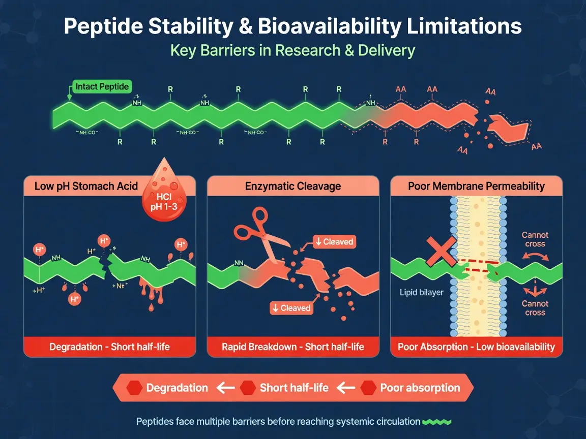
Why Do Peptide Stability and Bioavailability Limit Research Use?
Pharmacokinetics and delivery challenges remain central when investigators ask how do peptides work in vivo or in complex media. Peptides are susceptible to proteolytic cleavage by serum proteases, renal clearance, and hepatic metabolism. Their plasma half-life can be minutes to hours, depending on sequence and modifications. In laboratory research, these limitations are managed through structural modifications such as PEGylation, cyclization, D-amino acid substitution, or N-terminal capping.
PEGylation attaches polyethylene glycol chains to increase hydrodynamic radius and reduce renal filtration. Cyclization constrains the peptide backbone, improving proteolytic stability and sometimes receptor affinity. These modifications can change how do peptides work by altering binding kinetics or signal duration. Researchers must validate that any modified peptide retains the desired biological activity in their specific assay. For a research overview of contraindications and limitations, see Peptide Therapy Contraindications: A Research Overview.
How Do Peptides Work in Laboratory Research?
In laboratory research, how do peptides work is typically evaluated through in vitro assays that measure receptor binding affinity (Kd), functional activity (EC50/IC50), and downstream second messenger accumulation. Common methods include radioligand binding, fluorescence polarization, cAMP ELISA, calcium flux assays, and reporter gene assays. These readouts allow researchers to quantify potency and efficacy without making therapeutic claims.
A standard workflow includes reconstituting lyophilized peptide in an appropriate solvent, verifying concentration by UV absorbance or amino acid analysis, and then testing across a dose range. Controls should include vehicle, a reference agonist or antagonist, and a receptor-negative cell line where possible. The specific answer to how do peptides work depends on the receptor system, cell background, and assay endpoint. A plain-English explanation of these principles is available at How Peptides Work: A Plain-English Explainer.
💡 Key Takeaway: How Do Peptides Work in Laboratory Research?
Which Research Applications Rely on Peptide Mechanisms?
Common research applications of peptides include receptor pharmacology studies, structure-activity relationship (SAR) campaigns, cell signaling pathway mapping, and biomarker discovery. Peptides are also used as standards in mass spectrometry, as substrates in enzymatic assays, and as probes in imaging experiments. In each case, understanding how do peptides work at the molecular level informs experimental design and data interpretation.
For muscle-related research, investigators may examine peptide effects on myoblast proliferation or differentiation in vitro. A detailed protocol for muscle growth peptide research is available at 56. Muscle Growth Peptide Protocol. No anabolic or performance claims are made; all use remains research-only.
What Are the Best Storage Practices for Research Peptides?
Proper storage preserves peptide integrity and ensures reproducible results. Lyophilized peptides should be stored at -20°C or -80°C, protected from light and moisture. Avoid repeated freeze-thaw cycles of reconstituted solutions. For short-term use, reconstituted peptides may be kept at 4°C for a few days, but long-term storage should be as frozen aliquots. Always follow the COA and manufacturer recommendations for each specific peptide.
Storage conditions affect how do peptides work over time. Oxidation of methionine or cysteine residues, aggregation, and deamidation can reduce biological activity. Researchers should document lot numbers, reconstitution date, solvent, and storage temperature. A simple checklist includes:
- Store lyophilized peptide at -20°C or below in a desiccated container.
- Reconstitute with sterile, peptide-compatible solvent (e.g., water, PBS, or dilute acetic acid as indicated).
- Aliquot reconstituted peptide to avoid repeated freeze-thaw.
- Record purity and sequence verification from the COA before first use.
- Discard any solution showing turbidity, precipitation, or unexpected color change.
Where Can Researchers Source Verified Peptides?
Research-grade peptide services at Peptide Headlines provide peptides with documented purity, sequence confirmation, and COAs. All products are sold strictly for in vitro laboratory research use. Investigators should request analytical data and verify that the peptide meets the requirements of their specific assay. The question how do peptides work in your protocol begins with a well-characterized starting material.
Before ordering, confirm the peptide sequence, modification state (e.g., amidation, acetylation), salt form, and net peptide content. These details influence solubility, stability, and receptor binding. Reputable suppliers provide transparent batch records and responsive technical support.
💡 Key Takeaway: Where Can Researchers Source Verified Peptides?
FAQ
How do peptides work at the cellular level?
Peptides work by binding to specific receptors on the cell surface or inside the cell, inducing a conformational change that activates intracellular signaling cascades. These cascades involve second messengers such as cAMP, Ca2+, and IP3, ultimately altering gene expression, enzyme activity, or ion flow.
What determines whether a peptide binds to a particular receptor?
Receptor binding depends on the amino acid sequence, charge distribution, and three-dimensional shape of the peptide. Hydrogen bonds, hydrophobic contacts, and electrostatic interactions between peptide side chains and receptor residues determine affinity and selectivity. Even subtle differences in transmembrane residues can discriminate between related receptor subtypes.
Why are second messengers important for how do peptides work?
Second messengers amplify the initial receptor signal inside the cell. For example, cAMP activates protein kinase A, while IP3 releases Ca2+ from the endoplasmic reticulum and DAG activates protein kinase C. These molecules convert a single binding event into a robust cellular response.
How do researchers overcome peptide stability and delivery challenges?
Researchers use structural modifications such as PEGylation, cyclization, D-amino acid substitution, or N-terminal capping to improve proteolytic stability and reduce clearance. In vitro assays also use controlled media and protease inhibitors to maintain peptide integrity during experiments.
What should I check before using a research peptide in an assay?
Verify the certificate of analysis (COA) for HPLC purity, mass spectrometry sequence confirmation, and net peptide content. Check the storage conditions, reconstitution solvent, and lot-specific data. Proper handling and documentation ensure reproducible results.
Topics covered in this article:
Ready to Take the Next Step?
Every product includes lot-specific documentation and third-party purity testing.
Explore Our Research Products →Free shipping on qualifying orders · Batch COA on request